Lower Leg Bone Diagram - Lower Leg Muscle Diagram Blank Sketch Coloring Page. Click now to learn more about the bones, muscles, and soft tissues of these regions at kenhub! The lower leg is a major anatomical part of the skeletal system. Pelvic girdle—made up of the ilium, ischium, and pubic bone, which form a. Ankle and foot pain massage therapy connections. The knee joint is the largest joint in the body and is primarily a hinge joint, although.
Lower jaw (mandible) collar bone. Its presence is accepted but. Here's a diagram with the tibia bone labelled, as well as the fibula. Labeled images using 3d reconstructions and an angiographic view. Anterior view with primary bones names.
Posterior Calf Anatomy Muscles Of The Lower Leg Diagram Calf Muscles Diagram - Human | Leg ... from i.pinimg.com The knee is a strong but flexible hinge joint. They allow you to move and provide support for your upper body. The foot bones shown in this diagram are the talus, navicular, cuneiform, cuboid, metatarsals and calcaneus. Together with the upper leg, it forms the lower extremity. Cheek bone (zygoma) upper jaw (maxilla). The structure of the bone within the head and neck and the upper part of the shaft of the femur would do credit to an engineer who had worked out the (left) the radius and the ulna, bones of the forearm; Initially, the pain develops during the run but may even resolve during the course of the run. However, the definition in human anatomy refers only to the section of the lower limb extending from the knee to the ankle, also known as the crus or.
Proximally, there are five key features of the tibia:
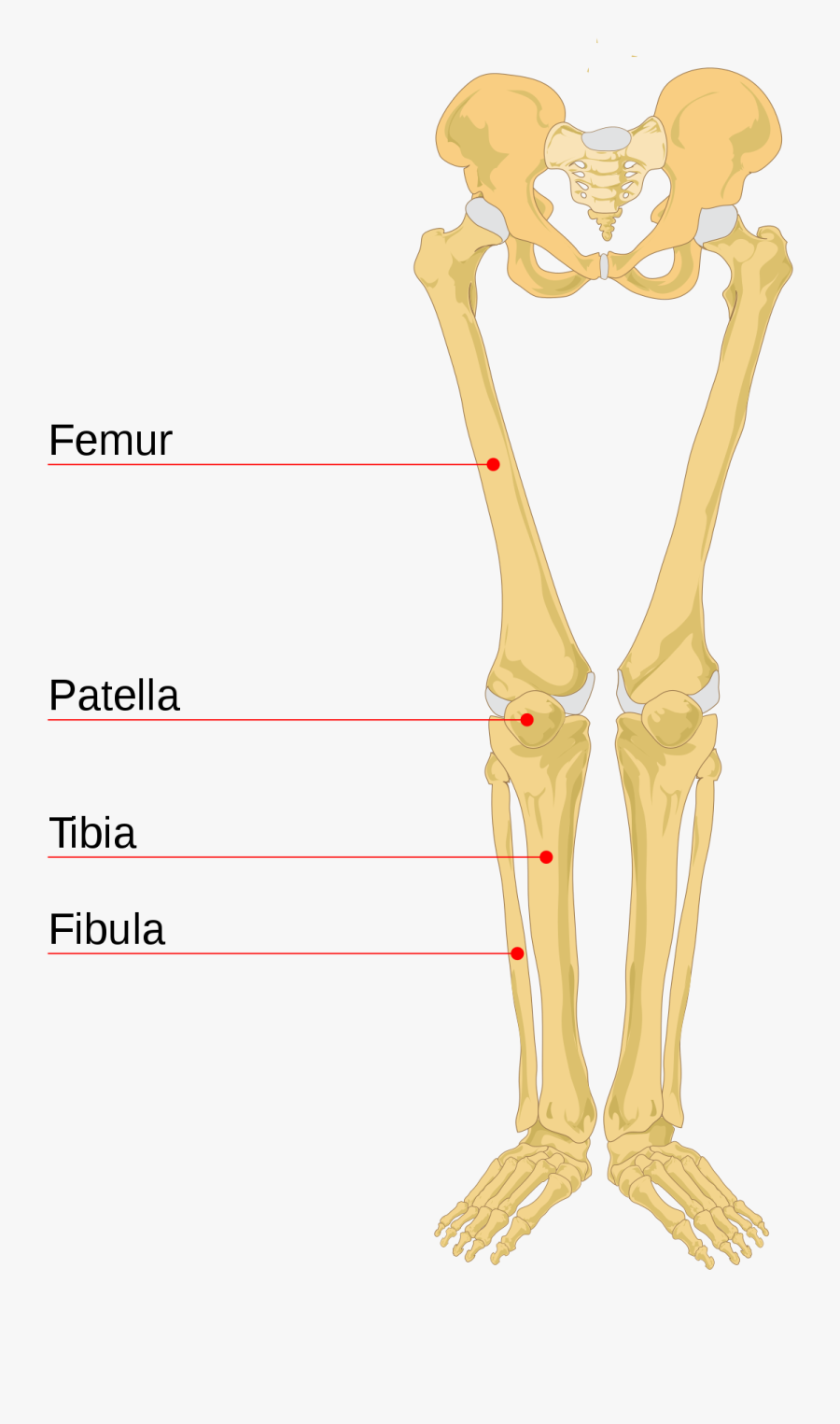
The tibia, or shin bone, spans the lower leg, articulating proximally with the femur and patella at the knee joint, and distally with the tarsal bones, to form the ankle joint. The typical presentation is bone pain with impact. Pelvic girdle—made up of the ilium, ischium, and pubic bone, which form a. The fibula is a long, skinny lower leg bone that looks rather fragile. Your leg bones are the longest and strongest bones in your body. Cited after worker's leg amputated. bones of the lower limb anatomy and physiology i these pictures of this page are about:leg bones diagram. Proximally, there are five key features of the tibia: It widens and forms two condyles. Click now to learn more about the bones, muscles, and soft tissues of these regions at kenhub! The thigh bone, or femur, is the large upper leg bone that connects the lower leg bones (knee joint) to the pelvic bone (hip joint). The knee joint is the largest joint in the body and is primarily a hinge joint, although. Bones of the leg and foot, lower leg bone anatomy, leg bones anatomy, leg muscles, leg bones diagram, leg bone structure, leg anatomy muscles, parts of the lower leg. They allow you to move and provide support for your upper body.
Find stockbilleder af lower leg bone anatomy anterior view i hd og millionvis af andre royaltyfri stockbilleder, illustrationer og vektorer i shutterstocks samling. Cheek bone (zygoma) upper jaw (maxilla). Download a free preview or high quality adobe illustrator ai, eps, pdf and high resolution jpeg versions. License image the bones of the leg are the femur, tibia, fibula and the foot bones shown in this diagram are the talus, navicular, cuneiform, cuboid, metatarsals and fibula, outer of two bones of the lower leg or hind limb. The fibula is a long, skinny lower leg bone that looks rather fragile.
Lower Limb Bones - The Human Skeletal System from sites.google.com The lower leg is comprised of two bones, the tibia and the smaller fibula. Structurally, bones are somewhat elastic because they are primarily made up of collagen. Cheek bone (zygoma) upper jaw (maxilla). The tibia (also called the shinbone) is located near the midline of. Here's a diagram with the tibia bone labelled, as well as the fibula. When you stand or walk, all the weight of your upper body rests on them. The tarsals are ankle bones and, along with the other bones in the foot (the metatarsals and phalanges), support weight and act as shock absorbers for the body. Diagram of lower leg bones posted on march 25, 2019 by admin this image shows the structure of tibia and fibula left panel legs bone diagram 20 13 asyaunited de u2022 hip drawing outline foot overview of bones the lower limb posterior and anterior view respectively 62 infographic diagram of.
This part of the interactive atlas of anatomy of the human body is about the arterial vasculature of the pelvic girdle, pelvis, thigh, knee, leg and foot and the.
Bones of the leg and foot, lower leg bone anatomy, leg bones anatomy, leg muscles, leg bones diagram, leg bone structure, leg anatomy muscles, parts of the lower leg. Labeled images using 3d reconstructions and an angiographic view. Your legs are two of your most important body parts. Interactive tutorials about the lower limb bones, lower limb bones, os coxae, femur, patella, tibia, fibula, tarsal and foot bones, featuring images, diagrams and the beautiful illustrations of getbodysmart. Its presence is accepted but. Anterior view with primary bones names. The bones of the leg are the femur, tibia, fibula and patella. You'll learn about the muscles, bones, and other structures of each area of the leg. The foot bones shown in this diagram are the talus, navicular, cuneiform, cuboid, metatarsals and calcaneus. The lower leg is comprised of two bones, the tibia and the smaller fibula. The tibia (also called the shinbone) is located near the midline of. It is situated on the lateral (or little toe) side of the leg. Leg length discrepancy (lld) or anisomelia, is defined as a condition in which the paired lower extremity limbs have a noticeably unequal length.
Together with the upper leg, it forms the lower extremity. The foot bones shown in this diagram are the talus, navicular, cuneiform, cuboid, metatarsals and calcaneus. Lower jaw (mandible) collar bone. The thigh bone, or femur, is the large upper leg bone that connects the lower leg bones (knee joint) to the pelvic bone (hip joint). Leg length discrepancy (lld) or anisomelia, is defined as a condition in which the paired lower extremity limbs have a noticeably unequal length.
Leg Bone Wikipedia Lower Leg Bones Diagram Legs Bones - Human Skeleton , Free Transparent ... from www.clipartkey.com The typical presentation is bone pain with impact. The knee is a strong but flexible hinge joint. The foot bones shown in this diagram are the talus, navicular, cuneiform, cuboid, metatarsals and calcaneus. The lower leg has a structure by two bones. It is situated on the lateral (or little toe) side of the leg. Most bones (particularly the long bones of the arms and legs — which make up the appendicular bones of the lower extremity. The primary cells in this area are termed as the calf. Master leg and knee anatomy using our topic page.
Your upper and lower leg are connected by a hinge joint.
Leg length discrepancy (lld) has been a controversial issue among researchers and clinicians for many years. The fibula is a long, skinny lower leg bone that looks rather fragile. Moreover, the fibula is the smaller bone that goes towards the back part of the leg. The foot bones shown in this diagram are the talus, navicular, cuneiform, cuboid, metatarsals and calcaneus. Master leg and knee anatomy using our topic page. Initially, the pain develops during the run but may even resolve during the course of the run. The lower leg is comprised of two bones, the tibia and the smaller fibula. Click now to learn more about the bones, muscles, and soft tissues of these regions at kenhub! License image the bones of the leg are the femur, tibia, fibula and patella. The knee is a strong but flexible hinge joint. The lower leg is a major anatomical part of the skeletal system. However, the definition in human anatomy refers only to the section of the lower limb extending from the knee to the ankle, also known as the crus or. Pelvic girdle—made up of the ilium, ischium, and pubic bone, which form a.
The lower leg is comprised of two bones, the tibia and the smaller fibula leg bone diagram. The largest and most medial leg bone, forming both the knee and ankle joints.
Share :
Post a Comment
for "Lower Leg Bone Diagram - Lower Leg Muscle Diagram Blank Sketch Coloring Page"


Post a Comment for "Lower Leg Bone Diagram - Lower Leg Muscle Diagram Blank Sketch Coloring Page"